THORAX
INTRODUCTION
Thorax (Latin chest) forms the upper part of the trunk of the body. It not only permits boarding and lodging of the thoracic viscera, but also provides necessary shelter to some of the abdominal viscera.
The trunk of the body is divided by the diaphragm into an upper part, called the thorax and a lower part, called the abdomen.
SURFACE LANDMARKS OF THORAX
BONY LANDMARKS
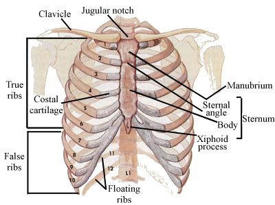
1. Suprasternal or jagular notch: It is felt just above the superior border of the manubrium between the sternal ends of the clavicles. The trachea can be palpated in this notch.
2. Sternal angle/angle of Luis: It is felt as a transverse ridge about 5 cm below the suprasternal notch.
3. Xiphisternal joint: The coastal margin on each side is formed by the seventh to tenth costal cartilages. The depression in the angle is also known as epigastric fossa. The xiphoid (GREEK sword) process lies in the floor of the epigastric fossa.
Fig.: Shape and construction of the thoracic cage as seen from the front
4. Costal cartilages: The second coastal(Latin rib) cartilage is attached to the sternal angle. The seventh cartilage bounds the upper part of the infrasternal angle.
5. Ribs: The scapula overlies the second to seventh ribs on the posterolateral aspect of the chest wall. The tenth rib is the lowest point, lies at the level of the third lumbar vertebra.
6. Thoracic vertebral spines: The first prominent spine felt at the lower part of the back of the neck is that of the seventh cervical vertebra or vertebra prominens.
SOFT TISSUE LANDMARKS
1. The nipple: The position of the nipple varies considerably in females, but in males it usually lies in the fourth intercostal space 10 cm from the midsternal line.
2. Apex beat: It is a visible and palpable cardiac impulse in the left fifth intercostal space 9 cm form the midsternal line, or medial to the midclavacular line.
3. Trachea: It is palpable in the suprasternal notch midway between the two clavicles.
Fig.: Shape and construction of the thoracic cage
Fig.: Shape and construction of thoracic cage as seen form behind
4. Midclavicular or mammary plane: It is a vertical plane passing through the midinguinal point, the tip of the ninth costal cartilage and middle of clavicle.
5. Midaxillary line: It passes vertically between the two folds of the axilla.
6. Scapular line: It passes vertically along the inferior angle of the scapula.
BONES OF THORAX
RIBS OR COSTAE
1.There are 12 ribs on each side forming the greater part of the thoracic skeleton. The number may be increased by development of a cervical or a lumber rib; or the number may be reduced to 11 by the absence of the twelfth rib.
2.The ribs are bony arches arranged one below the other. The gaps between the ribs are called intercostal spaces. The spaces are deeper in front than behind, and deeper between the upper than between the lower ribs.
3.The ribs are placed obliquely, the upper ribs being less oblique than the lower. The obliquity reaches its maximum at the ninth rib, and thereafter it gradually decreases to the twelfth rib.
4.The length of the ribs increases from the first to the seventh ribs, and then gradually decreases from the eighth to twelfth ribs.
5.The breadth of the ribs decreases from above downwards. In the upper ten ribs, the anterior ends are broader than the posterior ends.
6.The first 7 ribs which are connected through their cartilages to the sternum are called true ribs, or vertebrosternal ribs. The remaining five are false ribs. Out of these the cartilages of the eighth, ninth and tenth ribs are joined to the next higher cartilage and are known as vertebrochondral ribs. The anterior ends of the eleventh and twelfth ribs are free and are called floating ribs or vertebral ribs.
7. The first two and last three ribs have special features, and are atypical ribs. The third to ninth ribs are typical ribs.
Fig.: Posterior end of a typical rib
6.The first 7 ribs which are connected through their cartilages to the sternum are called true ribs, or vertebrosternal ribs. The remaining five are false ribs. Out of these the cartilages of the eighth, ninth and tenth ribs are joined to the next higher cartilage and are known as vertebrochondral ribs. The anterior ends of the eleventh and twelfth ribs are free and are called floating ribs or vertebral ribs.
7. The first two and last three ribs have special features, and are atypical ribs. The third to ninth ribs are typical ribs.
TYPICAL RIBS
1.The anterior end bears a concave depression. The posterior end bears a head, a neck and a tubercle.
2.The shift is convex upwards and there is a costal groove situated along the lower part of its inner surface, so that the lower border is thin and the upper border rounded.
Each rib has two ends, anterior and posterior. Its shaft comprises upper and lower borders and outer and inner surfaces.
The anterior sternal end is oval and concave for articulation with its costal cartilages.
OSSIFICATION OF A TYPICAL RIB
A typical rib ossifies in cartilage from:
a) One primary centre(for the shift) which appears, near the angle, at about the eighth week of intrauterine life.
b) Three secondary centres, one for the head and two for the tubercle, which appears at puberty and unite with the rest of the bone after 20 yrs.
First Rib
1.It is the shortest, broadest and most curved rib.
2.The shaft is not twisted. There is no costal groove.
3.It is flattened from above downwards so that it has superior and inferior surfaces; outer and inner borders.
Features of First Rib
1.The anterior end is larger and thicker than that in the other ribs. It is continuous with the first costal cartilage.
2.The posterior end comprises the following:
a)The head is small and rounded. It articulates with the body of first thoracic vertebra.
b)The neck is rounded directed laterally, upwards and backwards.
c
)The tubercle is large. It coincides with the angle of the rib. It articulates with the transverse process of first thoracic vertebra to form the costotransverse joint.
3.The shaft(body) has two surfaces, upper and lower and two borders, outer and inner.
a)The upper surface is marked by two shallow grooves, separated near the inner border by the scalene tubercle.
b)The lower surface is smooth and has no costal groove.
c)The outer border is convex, thick behind and thin in front.
d)The inner border is concave.
SECOND RIB
FEATURES
The features of the second rib are:
1.The length is twice that of the first rib.
2.The shaft is sharply curved,like that of the first rib.
3.The non-articular part of the tubercle is small.
4.The angle is slight and is situated close to the tubercle.
5.The shaft has no twist.The outer surface is convex and faces more upwards than outwards.Near its middle, it is marked by a large rough tubercle. This tubercle is a unique feature of the second rib. The inner surface of the shaft is smooth and concave. It faces more downwards than inwards. There is a short costal groove on the posterior part of this surface.
The posterior part of the upper border has distinct outer and inner lips. The part of the outer lip just in front of the angle is rough.
TENTH RIB
The tenth rib closely resembles a typical rib, but:
1.Shorter
2.Has only a single facet on the head, for the body of the tenth thoracic vertebra.
Eleventh and Twelfth Ribs
Eleventh and twelfth ribs are short. They have pointed ends.The necks and tubercles are absent.The angle and costal groove are poorly marked in the eleventh rib and are absent in the twelfth rib.
 |
| Fig.:Twelfth rib inner and outer surface |
COSTAL CARTILAGES
The costal cartilages represent the unossified anterior parts of the ribs. They are made up of hyaline cartilage. They contribute materially to the elasticity of the thoracic wall.
The medial ends of the costal cartilages of the first seven ribs are attached directly to the sternum. The eighth, ninth and tenth cartilages articulate with one another and form the costal margin. The cartilages of the eleventh and twelfth ribs are small. Their ends are free and lie in the muscles of the abdominal wall.
The direction of the costal cartilages is variable. As the first costal cartilage approaches the sternum, it descends a little. The second cartilage is horizontal. The third ascends slightly. The remaining costal cartilages are angular. They continue the downward course of the rib for some distance, and then turn upwards to reach either the sternum or the next higher costal cartilage. Each cartilage has two surfaces, anterior and posterior; two borders, superior and inferior; and two ends, lateral and medial.
STERNUM
The sternum is a flat bone, forming the anterior median part of the thoracic skeleton. In shape, it resembles a short sword. The upper part, corresponding to the handle, is called the manubrium. The middle part, resembling the blade is called the body. The lowest tapering part forming the point of the sword is the xiphoid process or xiphisternum.
The sternum is about 17 cm long. It is longer in males than in females.
Fig.: The sternum: Anterior aspect
Fig.: The sternum: Lateral aspect
MANUBRIUM
The manubrium is quadrilateral in shape. It is the thickest and strongest part of the sternum. It has two surfaces, anterior and posterior; and four borders, superior, inferior, and two lateral.
Body of the Sternum
The body is longer, narrower and thinner than the manubrium. It is widest close to its lower and opposite the articulation with the fifth costal cartilage. It has two surfaces, anterior and posterior; two lateral borders; and two ends, upper and lower.
XIPHOID PROCESS
The xiphoid process is the smallest part of the sternum. It is at first cartilaginous, but in the adult it becomes ossified near its upper end. It varies greatly in shape and may be bifid or perforated. It lies in the floor of the epigstric fossa.
Attachments
1.The anterior surface provides insertion to the medial fibres of the recctus abdominis, and to the aponeuroses of the external and internal oblique muscles of the abdomen.
2.The posterior surface gives origin to the diaphragm. It is related to the anterior surface of the liver.
3.The lateral borders of the xiphoid process give attachment to the aponeuroses of the internal oblique and transversus abdominis muscles.
4.The upper end forms a primary cartilaginous joint with the body of the sternum.
5.The lower end affords attachment to the linea alba.
DEVELOPMENT AND OSSIFICATION
The sternum develops by fusion of two sternal plates formed on either side of the midline. The fusion of two plates takes place in a craniocaudal direction.
Manubrium is ossified from 2 centres appearing in 5th month. Third and fourth sternebrae ossify from paired centres which appear in 5th and 6th months. These fuse with each other from below upwards during puberty Fusion is completed by 25 yrs of age.
Fig.: Ossification of sternum
VERTEBRAL COLUMN
The vertebral column is also called the spine, the spinal column, or back bone. It is the central axis of the body. It supports the body weight and transmits it to the ground through the lower limbs.
The vertebral column is made up of 33 vertebrae; 7 cervical, 12 thoracic, 5 lumbar, 5 sacral and 4 coccygeal. In the thoracic, lumbar and sacral regions, the number of vertebrae corresponds to the number of spinal nerves, each nerve lying below the corresponding vertebra. In the cervical region, there are eight nerves, the upper seven lying above the corresponding vertebrae and the eighth below the seventh vertebra. In the coccygeal region, there is only one coccygeal nerve.
Manubriosternal Joint
Manubriosternal joint is a secondary cartilaginous joint. It permits slight movements of the body of the sternumon the manubrium during respiration.
Costovertebral Joints
The head of a typical rib articulates with its own vertebra, and also with the body of the next higher vertebra, to form two plane synovial joints seperated by an intra-articular ligament. This ligament is attached to the ridge on the head of the rib and to the intervertebral disc.
Other ligaments of the joint include a capsular ligament and a triradiate ligament. The middle band of the triradiate ligaments forms the hypochordal bow, uniting the joints of the two sides.
Costotransverse Joints
The tubercle of a typical rib articulates with the facet on anterior surface of transverse process of the corresponding vertebra to form a synovial joint.
The capsular ligament is straightened by three costotransverse ligaments. The superior costotransverse ligament has two laminae which extend from the crest on the neck of the rib to the transverse process of the vertebra above. The inferior costotransverse ligament passes from the posterior surface of the neck to the transverse process of its own vertebra.
Costochondral Joints
Each rib is continuous anteriorly with its cartilage, to form a primary cartilaginous joint. No movements are permitted at these joints.
Chondrosternal Joints
The first chondrosternal joint is a primary cartilaginous joint, it does not permit any movement. This helps in the stability of the shoulder girdle and of the upper limb.
The second to seventh costal cartilages articulate with the sternum by synovial joints. Each joint has a single cavity except in the second joint where the cavity is divided into two parts. The joints are held together by the capsular and radiate ligaments.
Interchondral Joints
The fifth to ninth costal cartilages articulate with one another by synovial joints. The tenth cartilage is united to the ninth by fibrous tissue.
Intervertebral Joints
Adjoining vertebrae are connected to each other at five joints. There is a median joint between the vertebral bodies, and four joints- two on the right side and two on the left side- between the articular processes.The joints between the articular processes are plane synovial joints.


Comments
Post a Comment