Posts

HYDROCEPHALUS

Image
                                                     HYDROCEPHALUS Term ‘hydro’ meaning water and ‘cephalus’ referring to the head. Hydrocephalus is a condition in which an accumulation of cerebrospinal fluid (CSF) occurs within the cavities of the brain. CSF has 3 crucial functions in our body- 1.      It acts as a shock absorber for the brain and spinal cord 2.      It acts as a vehicle for transporting nutrients in the brain and removing waste. 3.      It flows between the cranium and spine to regulate changes in pressure within the brain. This accumulation of CSF causes increased pressure inside the skull. Hydrocephalus can occur due to birth defects or can be acquired later in life. The 4 types of hydrocephalous are communicating, non-communicating, ex-vacuo, and normal pressure. SYMPTOMS The signs and symptoms of hydrocephalus may vary by age of onset. Infants In infants CSF builds up in CNS, causing the signs and symptoms of hydrocephalus in infa
Image
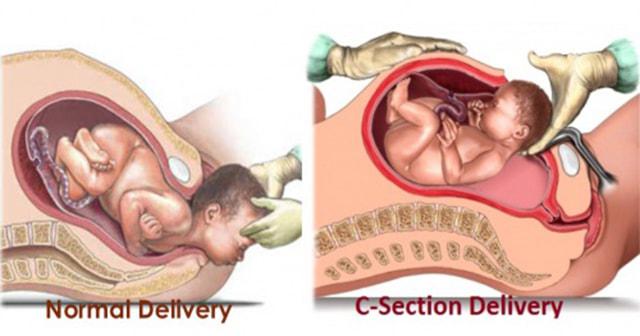
                                                  CAESAREAN SECTION Caesarean section, also known as C-section  or caesarean delivery, is the use of surgery to deliver one or more babies. A caesarean section is often necessary when a vaginal delivery would put the baby or mother at risk. This may include obstructed labour, twin pregnancy, high blood pressure in the mother, breech birth(bottom first instead of head first comes of the baby), or problems with the placenta or umbilical cord. A caesarean delivery may be performed based upon the shape of mother's pelvis or history of a previous C-section.                                         PROCEDURE  A C-section typically takes 45 minutes to an hour. It maybe done with a spinal block where the woman is awake or under general anesthesia.  A urinary catheter is used to drain the bladder and the skin of the abdomen is then cleaned with an antiseptic. An incision of about 15 cm (6 inches) is then made through the mot

HEART

Image
                                                        INTRODUCTION Mediastinum(plural-mediastina)(Latin intermediate ) is the middle space left in the thoracic cavity in between the lungs. Its most important content is the heart, enclosed in the pericardium in the middle part of the inferior mediastinum or the middle mediastinum. SUPERIOR AND INFERIOR MEDIASTINA For descriptive purpose, the mediastina is divided into superior mediastinum and the inferior mediastinum.                                                                            Fig.: Subdivisions of the mediastinum SUPERIOR MEDIASTINUM Boundaries Anteriorly: Manubrium sterni Posteriorly: Upper four thoracic vertebrae Superiorly: Plane of the thoracic inlet Inferiorly: An imaginary plane passing through the sternal angle in front, and the lower border of the body of the fourth thoracic vertebra behind. On each side: Mediastinal pleura Contents 1. Trachea and esophagus. 2. Muscles: Orig

THORAX

Image
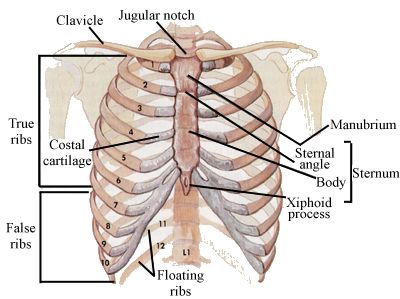
                                                     INTRODUCTION Thorax (Latin chest ) forms the upper part of the trunk of the body. It not only permits boarding and lodging of the thoracic viscera, but also provides necessary shelter to some of the abdominal viscera. The trunk of the body is divided by the diaphragm into an upper part, called the thorax  and a lower part, called the abdomen. SURFACE LANDMARKS OF THORAX BONY LANDMARKS 1.  Suprasternal or jagular notch : It is felt just above the superior border of the manubrium between the sternal ends of the clavicles. The trachea can be palpated in this notch. 2.  Sternal angle/angle of Luis : It is felt as a transverse ridge about 5 cm below the suprasternal notch. 3. Xiphisternal joint : The coastal margin on each side is formed by the seventh to tenth costal cartilages. The depression in the angle is  also known as epigastric fossa. The xiphoid (GREEK sword ) process lies in the floor of the epigastric fossa.