Posts

Showing posts from December, 2017

HEART

Image
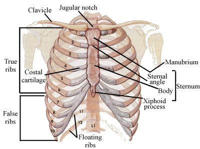
                                                        INTRODUCTION Mediastinum(plural-mediastina)(Latin intermediate ) is the middle space left in the thoracic cavity in between the lungs. Its most important content is the heart, enclosed in the pericardium in the middle part of the inferior mediastinum or the middle mediastinum. SUPERIOR AND INFERIOR MEDIASTINA For descriptive purpose, the mediastina is divided into superior mediastinum and the inferior mediastinum.                                                                            Fig.: Subdivisions of the mediastinum SUPERIOR MEDIASTINUM Boundaries Anteriorly: Manubrium sterni Posteriorly: Upper four thoracic vertebrae Superiorly: Plane of the thoracic inlet Inferiorly: An imaginary plane passing through the sternal angle in front, and the lower border of the body of the fourth thoracic vertebra behind. On each side: Mediastinal pleura Contents 1. Trachea and esophagus. 2. Muscles: Orig

THORAX

Image
                                                     INTRODUCTION Thorax (Latin chest ) forms the upper part of the trunk of the body. It not only permits boarding and lodging of the thoracic viscera, but also provides necessary shelter to some of the abdominal viscera. The trunk of the body is divided by the diaphragm into an upper part, called the thorax  and a lower part, called the abdomen. SURFACE LANDMARKS OF THORAX BONY LANDMARKS 1.  Suprasternal or jagular notch : It is felt just above the superior border of the manubrium between the sternal ends of the clavicles. The trachea can be palpated in this notch. 2.  Sternal angle/angle of Luis : It is felt as a transverse ridge about 5 cm below the suprasternal notch. 3. Xiphisternal joint : The coastal margin on each side is formed by the seventh to tenth costal cartilages. The depression in the angle is  also known as epigastric fossa. The xiphoid (GREEK sword ) process lies in the floor of the epigastric fossa.

Human Neural System

Image
CENTRAL NEURAL SYSTEM It comprises of the brain and spinal chord.                                   BRAIN The brain is the central information processing organ of our body. Brain lies in the cranium of the skull. Brain and Spinal cord are surrounded connective tissue membranes called as meninges. There are 3 meninges in humans; an outer layer called, dura mater , a very thin middle layer called arachanoid mater and an inner layer called pia mater . Human brain is divided into three parts - Fore brain( prosencephalon ), mid brain ( mesencephalon ) and hind brain ( rhombencephalon ).          FORE BRAIN It consists of olfactory lobes, cerebrum and diencephalon. Cerebrum  is the largest and most complex of all parts of human brain. A deep cleft divides the cerebrum longitudinally into two halves, which are termed as the left and right cerebral hemispheres are connected by a tract of nerve fibres called corpus callosum.