Human Neural System
CENTRAL NEURAL SYSTEM
It comprises of the brain and spinal chord.BRAIN
The brain is the central information processing organ of our body. Brain lies in the cranium of the skull. Brain and Spinal cord are surrounded connective tissue membranes called as meninges. There are 3 meninges in humans; an outer layer called, dura mater, a very thin middle layer called arachanoid mater and an inner layer called pia mater.
Human brain is divided into three parts - Fore brain(prosencephalon), mid brain (mesencephalon) and hind brain (rhombencephalon).
FORE BRAIN
It consists of olfactory lobes, cerebrum and diencephalon.
Cerebrum is the largest and most complex of all parts of human brain. A deep cleft divides the cerebrum longitudinally into two halves, which are termed as the left and right cerebral hemispheres are connected by a tract of nerve fibres called corpus callosum.

Fig.: Diagram showing sagittal section of the human brain


 |
| DISSECTION OF HUMAN SKULL |
The left part of the cerebrum controls the functions of right parts of the body while right part of cerebrum commands functions of left side of the body.
The interior of each cerebral hemisphere contains a lateral ventricle(=paracoel, first and second ventricle) filled with cerebrospinal fluid. The two ventricles open into third ventricle by a common aperture called foramen monro. Cerebrum consists of two parts- cerebral cortex and cerebral medulla.
The outer layer of cerebrum is called the cortex which comprises of many folds (usually elevated part) gyri and depression called suci.
Each cerebral hemisphere of the cerebrum is divided into four lobes: frontal,parietal,temporal and occipital lobes.
Table: Important areas present in the four lobes of cerebral hemisphere
Table: Important areas present in the four lobes of cerebral hemisphere

Diencephalon lies between cerebrum and mesencephalon.
Its cavity is called third ventricle or diocoel. It contains epithalamus, thalamus and hypothalamus.
Epithalamus is non-nervous part which is fused with piamater to form anterior choroid plexus. Behind it lies pineal body which is endocrine in function and secretes a hormone called melatonin. Epithalamus forms the roof of third ventricle.
Thalamus forms the lateral walls of the third ventricle and directs sensory impulses from lower parts of the brain and spinal cord to appropriate parts of the cerebrum. Just beneath the thalamus, hypothalamus forms the floor and the part of the lateral walls of the third ventricle.
Hypothalamus links nervous system to endocrine system (via hypothalamus-hypophyseal axis) and exercises a regulatory control on the functioning of endocrine glands by secreting neurohormones.
It contains higher centres of autonomic nervous system controlling hunger, thirst, sleep, fatigue, emotions, satisfaction, anger, pleasure, sexual behaviour, etc.
MID BRAIN
It is located between the thalamus/hypothalamus of the forebrain and pons of the hindbrain. A canal called the cerebral aqueduct passes through the midbrain. The dorsal portion of the midbrain consists mainly of four round swellings (lobes) called corpora quadrigemina. Corpora quadrgemina are equivalent to optic lobes of lower animals.
HIND BRAIN
Hind brain consists of cerebellum, medulla oblongata and pons varolii.
Cerebellum is the largest part of the hind brain consisting of two hemispheres and a median vermis. It is the centre for the maintenance of posture and equilibrium of the body and for the muscle tone. All activities of cerebellum are involuntary. They may, however, involve learning in early stages. Cerebellum is essential for controlling rapid muscular activities (e.g., running,typing talking).
The surface of cerebellum is also highly grooved. The grey matter of cerebellum is called cerebral cortex. A cross section of the hemisphere shows a branching tree like arrangement of grey and white matter called the arbor vitae("tree of life").
Medulla Oblongata, encloses a cavity called fourth ventricle. The medulla contains centres for controlling the functions of important organs, e.g., cardiac centre(heart), respiratory centre, vasomotor centre(for regulating diameter of blood vessels) and reflex centres(for swallowing,vomiting,peristalsis,secretions and activity of alimentary canal,salivation,coughing etc).
Pons varolli is situated in front of the cerebellum below the mid brain and above the medulla oblongata. It carries impulses from one hemisphere of the cerebellum to another. Functionally, the pons is concerned with maintenance of normal rhythm of respiration. It has got two respiratory centres- the pneumotaxic centre and apneustic centre.
The mid brain, pons varolii and medulla oblongata are collectively called the brain stem, connecting the fore brain and spinal cord.

Fig.: Median section of human brain
SPINAL CORD
The spinal coed is a tubular bundle of neural tissue that extends from the brain(medulla oblongata). In an adult the spinal cord is from 42 to 45 centimeters long. Its diameter varies at different level, being enlarged in the cervical and lumber regions. It begins in the atlas and tapers to a point, conus medullaris, in the first or second lumbar vertebra.
The spinal cord is formed of two types of nervous tissue: grey matter and white matter.
The grey matter is internal, i.e., surrounds the central canal. It has the form of letter H in cross section (also called butterfly-shaped). The arms of H are called posterior and anterior grey columns.
The regions of grey matter projecting towards the back of the body are called the dorsal horns, whereas those oriented towards the front are the ventral horns.
The grey matter is surrounded by white matter, which consists of groups of myelinated axons.
The spinal nerves tracts are divisible into two, ascending (conducting sensory impulses towards brain) and descending (conducts motor impulses from brain).
Spinal cord conducts impulses to and from the brain and controls most of the reflex activities.
PERIPHERAL NEURAL SYSTEM
The nerve running outside brain and spinal cord constitute peripheral neural system. These nerves are of two types: cranial nerves (nerves originating from brain) and spinal nerves (nerves originating from spinal cord).
CRANIAL NERVES
There are 12 pairs of cranial nerves, 10 pairs originate from the brain stem. They are numbered I to XII in Roman numerals.
SPINAL NERVES
Spinal nerves are 31 pairs of spinal nerves arising from either side of the spinal cord. Each spinal nerve is a mixed nerve, containing both sensory and motor nerve fibres running between the spinal cord and peripheral tissues.The nerve originates from dorsal(posterior) and ventral(anterior) nerve roots. The dorsal root has only nerve fibres and ventral root has only motor nerve fibres. Spinal nerves are classified into 5 groups-8 pairs of cervical nerves; 12 pairs of thoracic nerves; 5 pairs of lumbar nerves; 5 pairs of sacral nerves, and one pair of coccygeal.
REFLEX ACTION
Reflex action is an immediate involuntary action of any organ or part of the body in response to a particular stimulus. Reflex action was discovered by Marshall Hall(1833). The nervous pathway taken by nerve impulses in a reflex action is called reflex arc.
Mechanism of reflex action
For a reflex action five things are normally essential: receptor,sensory nerve fibres (afferent pathway), a part of the central neural system, motor nerve fibres(efferent pathway) and effector organ such as muscles and glands.
Reflexes are categorized into two : unconditioned and conditioned reflex. Unconditioned reflex is inborn, unlearned response to a stimulus or any change in the environment. Whereas, conditioned reflexes are not inborn but are acquired on past experience training or learning. It was demonstrated by I.P. Pavlov.
SENSORY RECEPTORS
Sensory receptors enable us to detect changes in our own body and objects and events in the word around us. Some receptors are simple and primitive type such as unspecialised sensory neuron whose terminal end is capable of detecting stimuli. For example, olfactory cells. The most complex sensory receptors consist of numerous sense cells, sensory neurons and associated accessory structures. For example, eye and ear.
EYE
The organ of sight are a pair of eyes in human. The eyes are situated in deep protective bony cavities, called the orbits or eye sockets of the skull. Eye consists of tissues present in three concentric layers:
- Outermost fibrous layer consists of sclera and cornea.
- Middle vascular layer (also called uvea) consists of choroid, ciliary body and iris.
- Inner most nervous layer consists of retina.
- Outermost fibrous layer consists of sclera and cornea.
- Middle vascular layer (also called uvea) consists of choroid, ciliary body and iris.
- Inner most nervous layer consists of retina.
FIBROUS LAYER
Sclera, an opaque outermost covering, maintains the shape of the eyeball and protects all the inner layers of the eye.
Cornea is a thin transparent, front part of sclera which lacks blood vessels but is rich in nerve endings. Cornea allows the light to pass into the eye. The cornea also serves as a filter, screening out some of most damaging ultraviolet (UV) wavelength in sunlight.
Conjunctiva is a thin transparent layer present over the cornea and is continuous with the skin over the eye. It protects the cornea and also secretes oils and mucous that moisten and lubricate the eye.
VASCULAR LAYER
Choroid is a pigmented layer present beneath the sclera. It contains numerous blood vessels and nourishes the retina.The choroid layer is thin over the posterior two-thirds of the eyeball, but it becomes thick in the anterior part to form the ciliary body.
Ciliary body holds the lens in the position, stretching and relaxation of this changes the focal length of the lens for accommodation.
Iris forms a pigmented circle of muscular diaphragm attached to the ciliary body in front of the lens. Its pigment gives eye its colour. The movement of iris controls the size of the pupil.
NERVOUS LAYER
The inner layer is the retina and it contains three layers of cells(from inside to outside)- ganglion cells, bipolar cells and photo receptor cells. The photoreceptors or visual cells are of two types: rod cells and cone cells, named after their shapes. Both have light- sensitive pigments. The daylight(photopic) vision and colour vision are functions of cones and the twilight (scotopic) vision is the function of the rods.
Optic nerves contains the fibres of the sensory neurons and leaves the eyeball from the back side. The point of departure of optic nerve through the retina does not have any rod or cones and thus produce a blind spot, there is a yellowish pigmented spot called macula lutea with a central pit called the fovea. The fovea is a thinned- out portion of the retina where only the cones are densely packed. It is the point where the visual acuity (resolution) is the greatest.
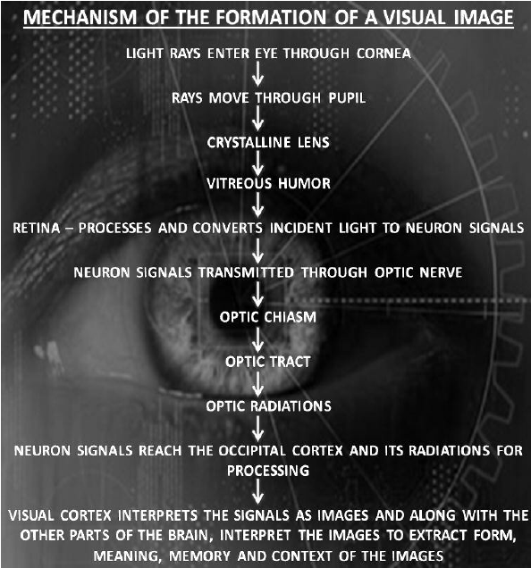
The light rays in visible wavelength, focussed on the retina through the cornea and lens, generate potentials (impulses) in rods and cones. The human eye is composed of Opsin (a protein) and retinal (an aldehyde of vitamin A).


MECHANISM OF VISION
Light induces dissociation of the retinal from opsin resulting in changes in the structure of the opsin. This causes membrane permeability changes. As a result, potential differences are generated in the photoreceptor cells. This produces a signal that generates action potential in the ganglion cells. These action potentials(impulses) are transmitted by the optic nerves to the visual cortex area of the brain.

EAR
The ears perform two sensory functions, hearing and maintenance of body balance. Anatomically, the ear can be divided into three major sections called the outer ear, the middle layer and the inner ear.
Outer ear comprises two parts, pinna and external auditory canal. Pinna is an oval, somewhat funnel- shaped skin covered flap of elastic cartilage and muscles. It collects sound waves and directs them into the external auditory canal. External auditory canal is an S-shaped tube leading inward from the pinna. The outer region of the canal bears hairs that serve to keep out the dust particles. Its inner region has oiled, tubular, apocrine ceruminous or wax glands, which lubricate and protect the lining of the meatus.
Middle ear is an air filled cavity in the temporal bone that opens via the auditory(Eustachian) tube into the nasopharynx. It includes tympanic membrane(or ear drum) and ear ossicles-the mealleus, the incus and the stapes. The malleus is attached to the tympanic membrane on one side and to the incus on other side. The incus in turn is connected with the stapes, which is connected with the stapes, which is attached to the oval membrane covering the oval window of the inner ear.
The function of the ossicles is to transmit and amplify sound waves across the tympanic cavity from the tympanic membrane to the oval window. The ossicles are connected in such a way as to act as a lever system to increase the force of the vibration from the ear drum.
Inner ear is delicate, irregular organ called membranous labyrinth. It is surrounded by an almost similarly shaped bony labyrinth. The membranous labyrinth is joined to the bony labyrinth at certain points, but its grater part is separated from the bony labyrinth by a narrow perilymphatic space. The space contains a watery fluid called perilymph. The membranous labyrinth is filled with another fluid called endolymph.
The coiled portion of the labyrinth is called cochlea. It is the main hearing organ. Internally it consists of three fluid-filled chambers or canals. The middle chamber called scala media is the most important canal or channel of the cochlea. It bears an upper membrane, the Reissner's membrane, and lower membrane, basilar membrane. On the basilar membrane a sensory ridge, the organ of corti is present. Organ of corti consists of hair cells(phonoreceptors), which bear 'hair' at the free surface and have synaptic contacts with the dendrites of neurons at the bases. The tips of 'hair' are embedded in a thin elastic membrane called tectorial membrane.

Fig.: T.S. Cochlea
The inner ear also contains a complex system called vestibular apparatus. The vestibular apparatus is responsible for maintenance of balance of the body and posture.
MECHANISM OF HEARING
The outer ear receives sound waves and directs them to the tympanic membrane. The tympanic membrane vibrates in response to the sound waves and these vibrations are transmitted through the ear ossicles to the oval window. The vibrations are passed through the oval window on to the fluid of the cochlea, where they generate waves in the lymph which induces ripple in the basilar membrane. These movements of the basilar membrane. As a result, nerve impulses are generated that are transmitted by the auditory nerves to the auditory cortex of the brain.

Fig.: Diagram showing the mechanism of hearing

Fig.: Flow chart of Mechanism of hearing
Comments
Post a Comment